Patented, proprietary antibody technology enables the SUCCEED Equine Fecal Blood Test to detect equine albumin and hemoglobin in a fecal sample, which can be indicative of pathological conditions of the GI tract, and can help distinguish between foregut and hindgut sources. This makes the SUCCEED FBT a powerful tool to aid the veterinarian’s diagnosis of GI tract conditions in horses.
Significant research went into understanding how antibodies could be used to differentiate between foregut and hindgut bleeding, developing the unique antibodies for equine albumin and hemoglobin, and testing their effectiveness. Review our antibody research to learn more about how these antibodies were developed and ELISA techniques used to test sensitivity and specificity.
Here’s how the antibody technology in the SUCCEED FBT works.
How Antibodies Become a Diagnostic Tool
Antibodies, also called “immunoglobulins,” are Y-shaped protein molecules secreted by specific white blood cells called plasma cells. Antibodies are an important part of human and animal immune systems because they identify and “flag” foreign objects, called antigens, in the body by attaching themselves. This allows other parts of the immune system to attack and destroy the foreign object, such as a virus or bacteria.
While antibodies are generally similar in structure, the tips – referred to as the antigen binding site – can vary widely. This allows for a large number of variations for binding different specific antigens. It is this variation in the antigen binding structures that allows for the production of antibodies specific to particular antigens which, in turn, makes antibody tests possible.
Antibodies Specific to Markers in Equine Blood
Two different antibodies are present in the membrane strips inside the FBT that are highly sensitive to specific protein markers in equine blood – one for albumin and one for hemoglobin. These blood markers act as antigens, thus reacting in the presence of the antibodies.
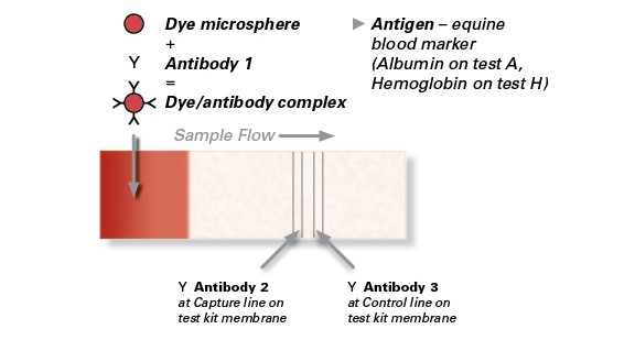
Each membrane strip in the SUCCEED FBT contains three different antibodies:
Y i. Antibody 1 specific for sample antigen (Albumin or Hemoglobin) is combined with a dye (dyed microspheres) and dried onto membrane
Y ii. Antibody 2 specific to the sample antigen applied to test strip at test line.
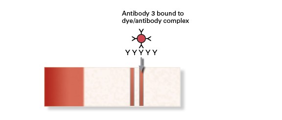
Y iii. Antibody 3 is a species-specific anti-immunoglobulin antibody that reacts to antibody 1 and is applied to strip at the control line.
What happens when applying fecal sample to the FBT well:
Two drops of a water/fecal sample mixture are applied to each well on the FBT test cassette. When you do this, the water/sample mixture wicks up the membrane strips inside the cassette, exposing the mixture to the antibodies. Here’s what happens:
Antibody 1 and dye mixture is rehydrated by sample and carried to capture and control lines.
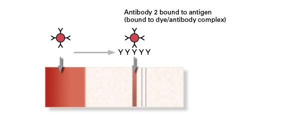
POSITIVE Test Result – antigen (blood marker) is present in sample
- Antigen binds to antibody 1 (with dyed microspheres) at one antigenic site…
- …then antigen binds to antibody 2 at capture line (carrying bound dyed microspheres)
- Dyed microspheres at capture line appear as a visible red line.
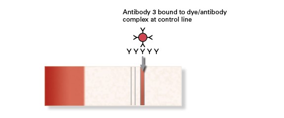
Control – confirms that test strip is valid
- Die/antibody complex are bound to antibody 3 at control line (whether antigen is present or not).
- Thus, a positive test result is indicated by the presence of TWO red lines on test strip.
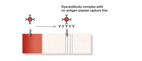
NEGATIVE Test Result
- Die/antibody complex is rehydrated and carried along membrane. As there is no antigen to bind to antibody 2 at the capture line, the die/antibody microspheres pass the capture line without being trapped.
- Die/antibody complex is bound by antibody 3 at the control line, causing a single red line to appear only at the control line.
